Is there X-ray radiation?
Ultrasound uses sound waves and no ionising radiation.

During salivary gland ultrasound a clinician moves a probe over the skin. Returning sound waves form an image that can show the location and structure of an abnormal area.
For training & practice ↓
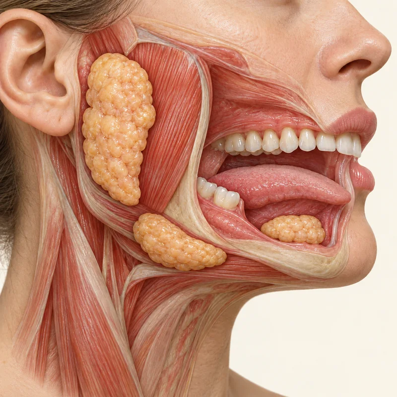
The clinician first locates the swelling. The parotid gland sits in front of the ear and the submandibular gland lies below the jaw.
Gel helps the probe make acoustic contact with the skin. Scanning in several directions shows sections of the gland and neighbouring structures.
If sampling is needed, a specialist can guide a needle towards the area using ultrasound. The laboratory examines the cells or tissue obtained.
The clinician first locates the swelling. The parotid gland sits in front of the ear and the submandibular gland lies below the jaw.
Gel helps the probe make acoustic contact with the skin. Scanning in several directions shows sections of the gland and neighbouring structures.
If sampling is needed, a specialist can guide a needle towards the area using ultrasound. The laboratory examines the cells or tissue obtained.
Ultrasound uses sound waves and no ionising radiation.
The standard scan is external. Sampling is a separate step discussed with you.
Additional imaging or sampling may be needed.
Three steps. No audio.
Each scene can also be selected manually.

Silent film with English on-screen text and simplified teaching images.
Ultrasound shows structures inside the body. Histology examines sampled tissue under the microscope.
Say when it started, whether it is growing and whether it swells around meals.
Glands, lymph nodes and other tissues are close together. Their position helps identify the origin of a lump.
Cells are collected to investigate further. The recommendation alone does not identify the lesion.
A question to bring along
Is imaging sufficient or do I need another test?
See a primary care doctor or specialist about a persistent swelling.
Choose an appointmentAppointment selection at DoctolibWhether a finding needs sampling depends on the clinical circumstances.
Editorial research: . General information, not an individual diagnosis or treatment recommendation. Teaching illustrations are simplified and do not depict a patient case.
If you turn this on, this website stores which guides you open, separately from advertising preferences. The markers expire after a fixed 30-day period from activation. The list is not sent to us by this feature. You can turn it off here and delete the markers.
Other people using the same browser profile can also see the markers. German and English versions share one marker. Expired entries are removed the next time you open a page with this feature; deleting browser data may remove them earlier. “Viewed” means opened, not read or completed.