Is a photograph a complete examination?
It shows only what is visible from that viewpoint.

During remote aligner monitoring, agreed photographs or image sequences are taken and sent to the treating team through its designated system. Images can help review visible changes and tray fit between visits. An unclear finding or new symptoms may require an in-person examination.
For training & practice ↓
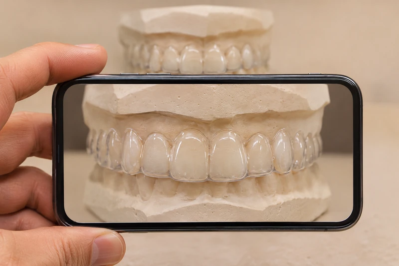
Even lighting, sharp focus and agreed views help comparison. The team specifies whether pictures are needed with the tray, without it or both.
A gap, damaged tray edge or missing attachment may be visible in suitable images. The images alone may not explain the underlying cause.
Report pain, a changed bite or persistent poor fit to your treating team. Examination and, where needed, a new scan can guide the next decision.
Even lighting, sharp focus and agreed views help comparison. The team specifies whether pictures are needed with the tray, without it or both.
A gap, damaged tray edge or missing attachment may be visible in suitable images. The images alone may not explain the underlying cause.
Report pain, a changed bite or persistent poor fit to your treating team. Examination and, where needed, a new scan can guide the next decision.
It shows only what is visible from that viewpoint.
Report new symptoms regardless of the photo schedule.
Use the channel designated by your treating team.
Three steps. No audio.
Each scene can also be selected manually.

Silent film with English on-screen text and simplified teaching images.
This describes targets and intended steps. Monitoring assesses the current state during treatment.
A randomised trial with 56 participants found fewer office visits in the remotely monitored group, but longer treatment on average. The need for a visit depends on your own progress.
Contact your treating team directly. An automated assessment cannot reliably determine causes of pain, tooth mobility or root changes.
A prospective study found differences in which teeth were flagged on repeat images. Ask if a message conflicts with what you notice. Follow the change rules agreed with your team.
A question to bring along
Who reviews the images and which changes should prompt a direct call?
Agree on a way to contact your own team about symptoms between scheduled visits.
Choose a dental appointmentAppointment selection at DoctolibA software message and a clinical decision serve different purposes.
Editorial research: . General information, not an individual diagnosis or treatment recommendation. Teaching illustrations are simplified and do not depict a patient case.
If you turn this on, this website stores which guides you open, separately from advertising preferences. The markers expire after a fixed 30-day period from activation. The list is not sent to us by this feature. You can turn it off here and delete the markers.
Other people using the same browser profile can also see the markers. German and English versions share one marker. Expired entries are removed the next time you open a page with this feature; deleting browser data may remove them earlier. “Viewed” means opened, not read or completed.